


New method brings Raman microscopy closer to detecting cancer stem-like cells
Researchers have developed a computational method that helps Raman micro spectroscopy distinguish molecular signals originating from cells from those produced by the materials surrounding them, improving the analysis of cells grown in complex biomaterial environments.
Raman microscopy can map the molecular composition of cells without fluorescent labels or staining. This makes it attractive for studying how cells respond to their surroundings. However, substrate such as glass, quartz and hydrogels also produce Raman signals, which can overlap with cellular information and vary across an image.
This is particularly important for hydrogels, which are increasingly used to mimic aspects of the cellular microenvironment. PAMPS-based hydrogels, for example, have previously been shown to promote cancer stem-like properties in several cancer models.
To address this problem, the researchers developed Adaptive Local Intensity Variation Adjustment (ALIVA), a computational method designed to recover cellular Raman signals while reducing contributions from the surrounding material. Rather than assuming that the background contribution is the same everywhere, ALIVA estimates it locally around each part of a cell using nearby cell-free measurements.
“It is like trying to listen to someone in a room where the surrounding noise changes depending on where you are standing,” says Professor Komatsuzaki. “Rather than removing the same noise everywhere, ALIVA estimates the local interference around each cell and corrects it accordingly.”
The team tested the method on Raman images of C2C12 cells carrying the SS18-SSX1 fusion gene, associated with synovial sarcoma, grown either on quartz or on a PAMPS hydrogel scaffold studied by Professor Tsuda and Tanaka, with a polymer scientist, Professor Gong in ICReDD. With conventional Raman preprocessing, cells measured on the two substrates appeared strongly different because much of the distinction originated from the substrate itself. After ALIVA correction, these differences were substantially reduced, and the cellular Raman spectra became much more pronounced and comparable.
The authors also investigated which molecular features a machine-learning algorithm used to distinguish cells grown on the two substrates. With conventional preprocessing, the algorithm relied largely on Raman features associated with the hydrogel. After ALIVA correction, features associated with the cells themselves, including cytochromes, proteins and phenylalanine, became much more important.
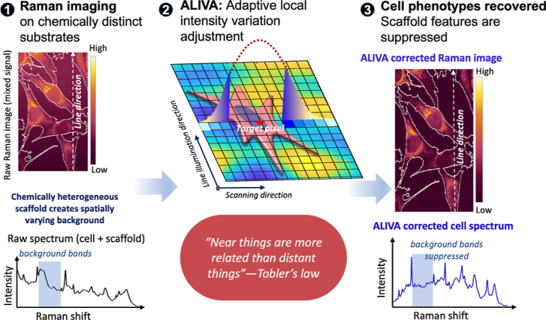
Figure. 1 Protocol of Adaptive Local Intensity Variation Adjustment (ALIVA)
Interestingly, after much of the surrounding-material contribution had been removed, the analysis revealed a subgroup of cells enriched among those grown on PAMPS hydrogel. The proportion of these cells was similar to stem-like populations reported previously in related hydrogel studies. Further experiments will be needed to determine whether this subgroup directly corresponds to a cancer stem-like phenotype.
The authors expect that ALIVA can make Raman imaging more reliable for studying how individual cells respond to complex biomaterial environments. In the future, the method supports investigations of cancer-cell plasticity, stem-like states and more complex biological systems such as organoids.
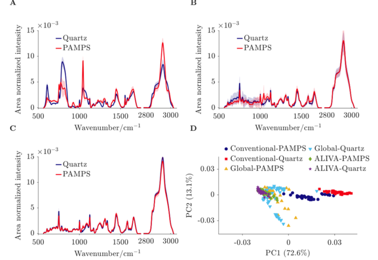
Figure. 2 Average and SD of Raman spectra of murine C2C12 SS18-SSX1 cells cultured on different scaffolds (quartz and PAMPS hydrogel), whose preprocessings are performed by (A) conventional, (B) global, and (C) ALIVA schemes. (D) PCA projections of average spectra from different scaffolds using the three different preprocessing schemes. The first two largest PCs cover 85.7% of the total variances of the spectral data over different scaffolds and three different preprocessings.
Contact
Professor Tamiki Komatsuzaki
Research Institute for Electronic Science, Hokkaido University
Tel/Fax: +81-11-706-9434
E-mail: tamiki[at]es.hokudai.ac.jp
Laboratory of Molecule & Life Nonlinear Sciences
URL https://mlns.es.hokudai.ac.jp/english.html

