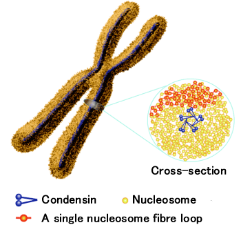


Professor Yoshinori Nishino (Laboratory of Coherent X-ray Optics, Research Institute for Electronic Science, Hokkaido University), Professor Kazuhiro Maeshima (National Institute of Genetics) and collaborators revealed by using strong X-rays from a large-scale synchrotron radiation facility SPring-8 that the DNA that constitutes the "blueprint of life" is irregularly bundled and stored "in a rather random fashion". The fine threads of DNA, with a diameter of 2 nanometers, are wrapped around spools made from proteins called histones to form nucleosome fibers that are about 11 nm in diameter. In 1976, the UK’s Klug (recipient of the Nobel Prize for Chemistry in 1982) and collaborators proposed that regular spiral folding of these nucleosome fibers could form a chromatin fiber of about 30-nm in diameter. A widely accepted and established theory about chromosome structure suggests that this chromatin fiber is spirally wrapped to form a fiber that is 100-nm in diameter, and subsequently 200- to 250-nm, then 500- to 750-nm fibers, and finally a regular and spiral hierarchy structure. In practice, the most famous textbook on molecular biology, "Molecular Biology of The Cell," and high-school biology textbooks describe this established theory.
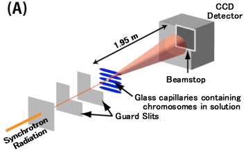
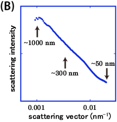
The research group has obtained results that disprove this established theory. They have found that the peak that appears in X-ray scattering data, which has been long regarded as a strong evidence for the chromatin fiber with a diameter of about 30 nm, is caused not by chromosomes themselves but by many ribosomes adhering to the surface of chromosomes. In addition, an ultra-small angle X-ray scattering instruments that was developed by Nishino et al., has allowed us to measure X-ray scattering up to the entire length scale of a 1-micrometer chromosome, although such large length-scale had been difficult to access. As a result, they found that there was no scattering peaks that would correspond to fibers of about 100-nm or about 200- to 250-nm predicted by this established theory. A series of these results points to the absence of the chromatin fiber and of higher-order structures in which the chromatin fiber is regularly bundled, despite the model of this theory.
The research was performed as a collaboration with Dr. Kazuhiro Maeshima, Professor of the National Institute of Genetics, Dr. Tetsuya Ishikawa, Chief Scientist of the Riken Harima Institute, Dr. Yasumasa Jouchi, Researcher (currently at the Japan Synchrotron Radiation Research Institute), Dr. Kazuki Ito, Contract Researcher (currently at Rigaku Corporation), Dr. Yukio Takahashi, Special Postdoctoral Researchers (currently at Osaka University), Dr. Naoko Imamoto, Chief Scientist of the Riken Advanced Science Institute, Dr. Hideaki Takata, JSPS Research Fellow at the National Institute of Genetics (currently at Osaka University), Saera Hihara (Graduate University for Advanced Studies), and Dr. Mikhail Eltsov and Dr. Achilleas Frangakis, EMBL, Germany. The research was supported by Promotion of X-ray Free Electron Laser Research of the Ministry of Education, Culture, Sports, Science and Technology (MEXT), JST’s CREST "Development and applications of scanning/transmission X-ray microscopy", and MEXT’s Grant-in-Aid for Scientific Research on Innovative Area "Physicochemical Field for Genetic Activities" and "Spying Minority in Biological Phenomena".
This work was published in the ‘EMBO Journal’ on April 4 (in the electronic version on February 17). "Human mitotic chromosomes consist predominantly of irregularly folded nucleosome fibres without a 30-nm chromatin structure", Y. Nishino, M. Eltsov, Y. Joti, K. Ito, H. Takata, Y. Takahashi, S. Hihara, A.S Frangakis, N. Imamoto, T. Ishikawa, and K. Maeshima, The EMBO Journal 31, 1644-1653 (2012).