


![]()
October 16th, 2023.
Abstract.
The research groups by Professor NEMOTO Tomomi* and Assistant Professor TSUTSUMI Motosuke* of National Institute for Physiological Sciences, National Institute of Natural Sciences and Dr. KOBAYASHI Kentaro at Division of Technical staffs of Research Institute of Electronic Science, Hokkaido University succeeded visualizing the nanoscale morphology in deep region on living brain applying for super-resolution radial fluctuation (SRRF) to the images acquired by two-photon microscopy. Since SRRF is a super-resolution technique based on image analysis, researchers can easily apply to fluorescence image and acquire images with a spatial resolution comparable to existing super-resolution microscopes for in vivo deep imaging.
*Professor Nemoto and Dr Tsutsumi belonged in RIES until September 2019.
Point.
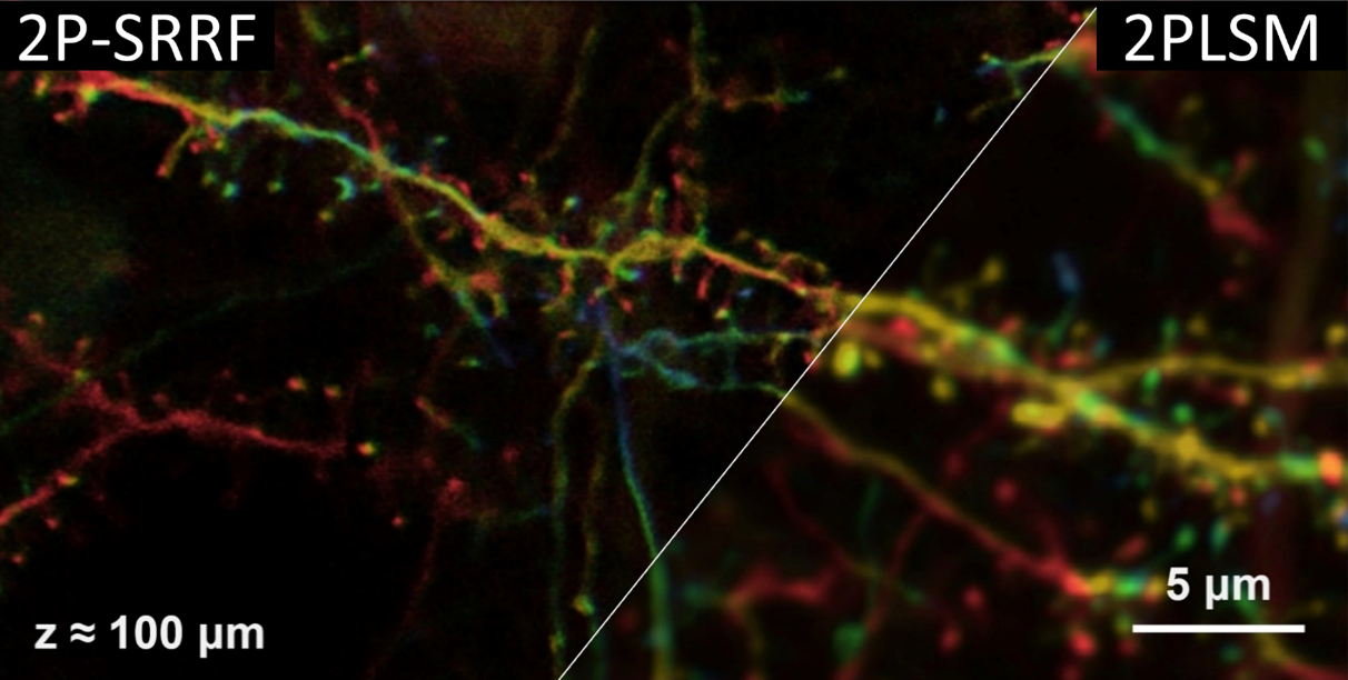
- It is still difficult to apply super-resolution microscopy for deep imaging due to the deterioration of light convergence properties in thick specimens. In this research, we succeeded in visualizing the nanostructures of neurons at 500 um depth in mouse brain in vivo.
- We applied super-resolution radial fluctuation (SRRF) to two-photon microscopy (2P-SRRF). We have successfully demonstrated that it is possible to observe tiny structures with spatial resolution beyond the diffraction limit of light in deep biological tissues.
- Researchers using existing two-photon microscopes may promptly apply the 2P-SRRF technique to expand the range of possible applications for super-resolution observation to deeper areas.
Original Research Article.
Motosuke Tsutsumi, Taiga Takahashi, Kentaro Kobayashi, Tomomi Nemoto* (* Corresponding Author), Frontiers in Cellular Neuroscience, 2023. DOI: 10.3389/fncel.2023.1243633